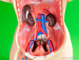
 Retroperitoneal tumors can be Benign or Malignant.
Solid tumors are commonly malignant (85%) and Cystic tumors are usually benign.
Types of Retroperitoneal tumors
Retroperitoneal Sarcoma - It is the most common type of malignant tumor.
Various types of Retroperitoneal sarcomas are:
- Liposarcoma
- Leiomyosarcoma
- Fibrosarcoma
- Malignant fibrous histiocytoma
In children, Rhabdomyosarcoma is the most common one.
Risk Factors:
Retroperitoneal tumors may be associated with
- Radiation
- Exposures to chemicals like vinyl chloride/thorium dioxide,
- Genetic Factors: Familial Gardner's syndrome, Familial retinoblastoma or Familial neurofibromatosis, Li Fraumeni syndrome, Germline mutations of p53 in chromosome 17.
Tumor characteristics
- Tumor arises from fat, areolar tissue, vessels, nerves, skeletal and smooth muscle, fascia, lymph node and lymphatic systems and tissues of embryologic origin.
- Sarcomas can be commonly solid, cystic or mixed. It spreads along the fascial planes enveloping the organs.
- Infiltration of organs occurs only at late stage.
- Spread of retroperitoneal sarcoma mainly occurs through blood to lungs and often to liver. Nodal spread occurs rarely (5%).
- Malignant tumors of the retroperitoneum are 4 times common than benign one.
Retroperitoneum is the 2nd most common site of malignant mesenchymal tumors first site is being lower extremities.
Clinical Features
 Most patients have initial asymptomatic course with eventual presentation as mass abdomen, abdominal pain and discomfort.
- Lump Abdomen (90%)
- Pelvic mass
- Vague abdominal pain and discomfort
- Anorexia, fatigue
- Vomiting
- Weight loss
- Fullness in abdomen
- Back pain due to compression over lumbar and pelvic nerves
- Leg pain
- Recurrent episodes of fever
Other presentations are —
- GI bleeding; intestinal/urinary obstruction
- Neurological manifestations by compression or invasion of neurological systems by tumor
- Iliac vein or IVC compression can cause lower limb varicosities, varicocele, and dilated abdominal veins with venous flow from below upwards, and oedema.
Investigations:
-
CT abdomen — Contrast CT abdomen (commonly used) and pelvis and CT chest (to see metastases) are essential investigations
-
CT-guided core biopsy
FNAC has got limited role.
-
MRI abdomen – identifies extent, desmoplastic reaction.
-
AFP, HCG and other tumour markers are done often.
-
Immuno-histochemistry is essential in many suspicious types of retroperitoneal tumors.
Treatment
-
Surgery is the main modality of therapy with wide excision— Complete en block excision is the ideal treatment
-
Limited resection; debulking are other options. Often it needs extensive surgical dissection; resection of adjacent bowel.
-
Postoperative chemotherapy and radiotherapy is used as adjuvant method
Radiotherapy - External beam RT (5000 cGy); or IORT (intraoperative RT during surgical resection) which reduces local recurrence and radiation enteritis.
Mesna, Adriamycin (main drug), ifosfamide and dacarbazine (MAID) are the chemotherapeutic agents used.
-
Recurrent disease needs re-exploration and to do complete clearance.
Prognosis
-
Prognosis depends on:
- Tumor grade—low or high
- Resectability
- Invasion into organs, veins, nerves
- Size of tumor < 5 cm or > 5 cm or > 10 cm
- Deep location
- Non liposarcoma in histology—poor prognosis
- Recurrent disease
- Distant spread
Tumor grade and completeness of resection without distant spread are important prognostic factors.
RP tumors has got overall 40% recurrence rate.
5-year survival for well-differentiated tumor is 75%; < 40% for undifferentiated tumors.

