13A, Pocket A, Phase 3, Ashok Vihar, Opposite Madrina Restaurant, Near Satyawati College, Delhi- 52

Liver Cancer

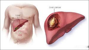
Liver is the most common site for metastasis from tumors of other organ system. Among the various types of liver tumors, Hepatocellular Carcinoma is the most common primary liver tumor.

Types of Liver tumors:

Benign Tumors:

  1. Liver cell adenoma
  2. Focal Nodular Hyperplasia
  3. Hemangioma
  4. Other tumors- Mesenchymal hamartomas, Fibrous tumors

Malignant Tumors:


  1. Hepatocellular Carcinoma
  2. Fibrolamellar HCC
  3. Intrahepatic Cholangiocarcinoma
  4. Other tumors - Hepatoblastoma, Sarcomas, Non Hodgkin's Lymphoma

Secondary Liver tumors


  1. Colorectal Metastasis
  2. Neuroendocrine metastasis
  3. Other tumor metastasis- Breast, Lung, Soft tissue sarcoma, upper GI tract tumors etc.

Hepatocellular Carcinoma (HCC)


  1. HCC is a primary malignant tumor of the liver with hepatocellular differentiation.
  2. Its incidence is increasing and is becoming 4th most common malignancy worldwide.
  3. HCC is the commonest primary liver malignancy (90%). But in the liver, secondaries is the most common malignancy (20 times more common than primary).
  4. HCC is common in cirrhotics and hepatitis B and hepatitis C virus infection.
  5. It is more common in Asian countries with a Male to female ratio of 4:1.
  6. Right lobe is commonly involved.

Risk Factors


  1. Hepatitis B and hepatitis C virus infection – more common in individuals who have chronic positive status for HBs Ag and chronic carriers. Vertical transmission of virus at birth raises HCC rate
  2. Alcoholic cirrhosis
  3. Aflatoxin B1, a product of fungus aspergillus
  4. Clonorchis sinensis infestation
  5. Smoking
  6. Haemochromatosis, α1 antitrypsin deficiency
  7. Hepatic adenoma—potentially malignant
  8. Environment related chemicals like DDT, nitrite and nitrate related food products; trichloroethylene (dry cleaning solvents), biphenyls, carbon tetrachloride, herbicides
  9. Anabolic steroids, polyvinyl chloride

Clinical Presentation


  1. Asymptomatic – Incidentally identified

  2. Symptomatic – Abdominal pain, weight and appetite loss, jaundice, mass/lump in abdomen

  3. Symptoms due to complications – Bleeding HCC can cause life threatening spontaneous haemoperitoneum after an attempt of biopsy;
    Jaundice (in 10% of HCC) due to obstruction by tumor of main intrahepatic bile ducts or CHD at porta hepatis or by tumor infiltration of biliary system

  4. Symptomatic due to metastasis – from lung metastases, bone metastases

  5. Paraneoplastic syndrome (1%) – hypoglycemia, hyperthyroidism etc.

Investigations


  1. Ultrasound Abdomen – Ultrasound shows a hyperechoic mass; Extent of tumor and tumor thrombi extension can be made out.

  2. Contrast Enhanced CT abdomen (CECT) - CT scan helps in assessment of size, location and extent, vascularity, portal vein invasion, nodal status, portal vein thrombosis. It helps to assess operability and plan for surgical resection.

  3. Tumor Markers - AFP (Alpha- Feto protein) >100 is suggestive of HCC. AFP>400 in >70% of HCC patients
    Other tumor markers- PIVKA II, DCP, Glypican-3

  4. Liver Function tests- Serum Bilirubin, Albumin, Liver enzymes (ALP, AST, ALT) and Prothrombin time.

  5. MRI abdomen - T2 weighted studies are useful for small HCC. MR angiography is also done to see tumor thrombus in portal vein, hepatic vein and IVC.

  6. Investigations in relation to hepatitis B and hepatitis C virus infections.

  7. Metastatic work up — HRCT of chest is essential
    FDG – PET scan and bone scans are useful in detecting the early metastatic diseases

Treatment

Definitive Treatment :
Surgery :


  1. When limited to one lobe, hemihepatectomy is done (Removal of upto 70% liver is compatible with life).
  2. Surgery is done only for Child's grade A and favourable Child's B group patients.
  3. It is the treatment of choice for operable HCC in non-cirrhotic patients.
  4. In cirrhotic patients (Child C, unfavourable Child B) with HCC, Total hepatectomy with orthotopic liver transplantation is required
  5. To bridge the waiting period for transplantation Radiofrequency ablation (RFA), Transarterial Embolization (TACE, TARE), ethanol/acetic acid injections are used.

Adjuvant Therapy


  1. Chemotherapy is given after surgery in cases with lymph nodes positive disease.
  2. Neoadjuvant (preoperative) chemotherapy also practiced to increase the operability/resectability of the tumor.
  3. Sorafenib (Nexavar) has been approved as targeted therapy for people with liver cancer. It is helpful for patients who are not a candidate for liver resection or liver transplant

Palliative Treatment


  1. Radiofrequency Ablation (RFA):
    It is thermal ablation of the tumor by passing 18G needle into the middle of the tumor and passing electric current of 500 kHz. This causes adequate cyto-destruction of the tumor.
    It can be done percutaneously under Ultrasound or CT guidance or through laparoscopy. Tumor less than 3 cm, tumor deep in parenchyma, tumor away from hilum is ideal for radioablation.

  2. Transarterial Embolization:
    Chemotherapeutic agents (TACE) or Radioisotopes (TARE) can be delivered via hepatic artery which is the sole blood supply to the tumor, thus causing significant dosage of these agents delivered to the tumor leading to more effective killing of the tumor cells.

  3. Percutaneous Ethanol or acetic acid injection

  4. Microwave Ablation or Cryoablation: Effects of the procedures are similar to RFA

© Copyright 2020, dr. pradeep jain. All rights reserved.